The short answer
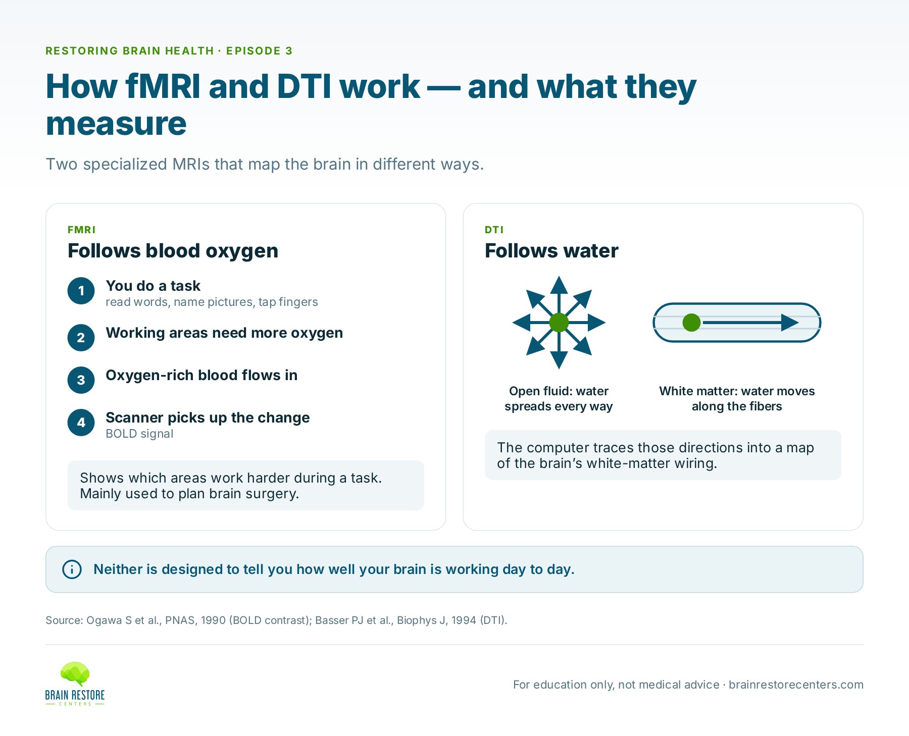
Not in the everyday sense. A functional MRI (fMRI) tracks changes in blood oxygen to show which areas of the brain work harder during a specific task, and it’s mainly used to plan brain surgery. DTI follows the movement of water to map the brain’s white-matter wiring. Both are powerful mapping tools, but neither is designed to tell you how well your brain is working day to day.
The names are confusing. “Functional MRI” sounds like exactly the test you’d want if you’re struggling and your standard brain MRI was clear. On Episode 3 of Restoring Brain Health, neurosurgeon Dr. Sharon Webb said she gets asked about these “special MRIs” all the time. Here’s how they actually work, and where they fit.
How does a functional MRI work?
Brain cells that are working harder need more oxygen, so the body sends extra blood to that area within a few seconds. Oxygen-rich and oxygen-poor blood behave slightly differently in a magnetic field. In 1990, researcher Seiji Ogawa and colleagues showed that an MRI scanner can pick up this difference, which they called BOLD contrast (blood oxygen level dependent). That discovery is the basis of every fMRI today.
During an fMRI you lie in the scanner and do simple tasks on cue: read words, name pictures, tap your fingers. Then you rest. A computer compares the “task” moments with the “rest” moments, and the areas with a stronger signal during the task are colored in. That’s the “lighting up” you see in news stories.
“It’s looking at what part of the brain is getting more blood flow while you’re doing certain tasks,” Dr. Webb says. It isn’t measuring how well your brain works overall.
What is a functional MRI actually used for?
Its main clinical job is surgical planning. Language and movement areas sit in roughly the same place in most people, but not exactly. If a tumor is close to the language center or the motor strip, the surgical team needs to know where that person’s areas actually are. A review from Harvard and Brigham and Women’s Hospital describes how fMRI is increasingly used to counsel patients, plan surgery and guide tumor removal near these critical areas.
In research, fMRI has taught scientists a great deal about how brain networks behave in large groups of people. But a single fMRI isn’t a standard way to rate one person’s day-to-day brain function, and it’s found mostly at academic medical centers.
How does diffusion tensor imaging (DTI) work?
DTI is another specialized MRI. Instead of blood, it follows water. Water molecules in the brain are always jiggling around. In open fluid they drift equally in every direction. But inside the brain’s white matter, water is hemmed in by long, insulated nerve fibers, so it moves more easily along the fiber than across it, like water running down a garden hose.
In 1994, Peter Basser and colleagues at the National Institutes of Health showed how MRI can measure this direction-dependent movement of water and use it to work out which way the fibers run. Computers can then trace those directions into colorful pictures of the brain’s wiring, called tractography. A common DTI number, fractional anisotropy (FA), describes how strongly water prefers one direction. Healthy, organized fibers usually have higher FA.
What is DTI used for, and can it show a brain injury?
Like fMRI, DTI is used mostly for surgical planning. Surgeons want to avoid cutting through important deep pathways, and DTI shows where they run.
DTI can also show diffuse axonal injury, which is widespread stretching or tearing of nerve fibers, when that injury is significant. Dr. Webb notes that a standard MRI can show it too if it’s bad enough. With milder injury, she says, “neither the DTI or your normal MRI is going to pick it up. You’re still going to have a functional issue, though, in your exam.”
Researchers are actively studying this. A 2012 meta-analysis that pooled 13 DTI studies found white-matter changes in the corpus callosum, the bridge between the brain’s two halves, when groups of people with mild brain injury were compared with groups without injury. That’s valuable science. But a difference between groups is not the same as a clear answer for one person sitting in a clinic, which is why the exam still matters so much.
“These aren’t functional tests. They’re not looking at the function of the brain.”
— Dr. Sharon Webb, on Restoring Brain Health, Episode 3
If DTI confirms an injury, what then?
Dr. Trevor Berry, who works with many people after brain injury, calls DTI “an amazing test.” In legal cases especially, it can provide validation that someone had a traumatic brain injury. But that only answers the first question.
“It’s a good starting point of validation, a great test, but you still got to paint the rest of the picture doing the thorough exam.”
— Dr. Trevor Berry, on Restoring Brain Health, Episode 3
Knowing that an injury happened doesn’t tell you which systems are affected now: balance, eye movements, memory, mood, sleep. That takes a functional look.
fMRI vs. DTI vs. functional assessment at a glance
- fMRI answers: where does blood flow increase during this task? Best for: mapping language and motor areas before surgery.
- DTI answers: where do the white-matter pathways run, and is there visible injury to them? Best for: surgical planning and showing more significant axonal injury.
- Functional assessment answers: how are this person’s brain networks working right now? Best for: a baseline when symptoms continue, measured again over time.
What actually measures how well your brain is working?
A functional picture is built from several sources that are read together:
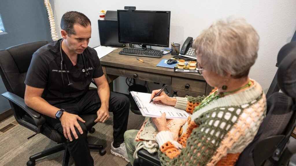
- A careful history and hands-on neurological exam
- A qEEG brain map, compared against normative data
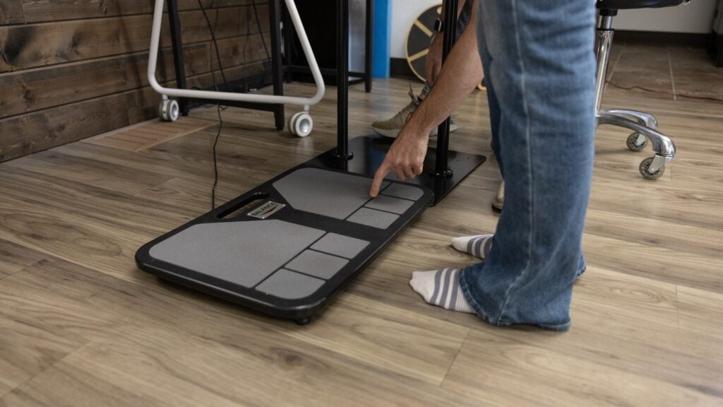
- Computerized balance testing on a force plate
- Eye movement, coordination, strength, and memory and thinking checks
At Brain Restore Centers, when these tests show areas of the brain not working together as well as they should, we call it network interference. Network interference is what we call it when testing shows that different areas and networks of the brain aren’t functioning and working together as well as they should. It’s something we measure, not a diagnosis.
Why several tests beat one is explained in why one brain test isn’t enough. For the full comparison table, see the photo and the video: what brain imaging shows, and what it can’t.
Frequently asked questions
Can I ask my doctor for a functional MRI to check my brain function?
You can ask, but fMRI is usually ordered for a specific reason, most often before surgery, and it’s mostly available at academic medical centers. It isn’t a routine test of overall brain function.
Is DTI the same as a regular MRI?
It’s done in the same scanner, with extra sequences and special computer processing. A regular MRI shows the shape of brain tissue; DTI estimates how water moves along the white-matter fibers.
Is a qEEG brain map the same as an fMRI?
No. An fMRI tracks blood oxygen during a task. A qEEG records the brain’s electrical activity directly from the scalp and compares it with normative data. They measure different things in different ways.
Studies referenced
- Ogawa S, Lee TM, Kay AR, Tank DW. Brain magnetic resonance imaging with contrast dependent on blood oxygenation. Proc Natl Acad Sci USA. 1990;87(24):9868-9872. https://pubmed.ncbi.nlm.nih.gov/2124706/
- Silva MA, See AP, Essayed WI, Golby AJ, Tie Y. Challenges and techniques for presurgical brain mapping with functional MRI. Neuroimage Clin. 2018;17:794-803. https://pubmed.ncbi.nlm.nih.gov/29270359/
- Basser PJ, Mattiello J, LeBihan D. MR diffusion tensor spectroscopy and imaging. Biophys J. 1994;66(1):259-267. https://pubmed.ncbi.nlm.nih.gov/8130344/
- Aoki Y, Inokuchi R, Gunshin M, Yahagi N, Suwa H. Diffusion tensor imaging studies of mild traumatic brain injury: a meta-analysis. J Neurol Neurosurg Psychiatry. 2012;83(9):870-876. https://pubmed.ncbi.nlm.nih.gov/22797288/
Watch the full conversation: Restoring Brain Health, Episode 3.
Find out how your brain is working right now
The first step is a Complimentary Brain Scan at a Brain Restore Center near you: a functional look at how your brain is working today. No pressure, no assumptions. If it looks like we may be able to help, we’ll talk about next best steps.
This article is for education and is not medical advice. It does not replace care from your own doctor. If you have sudden or severe symptoms, such as a sudden severe headache, new weakness or numbness, trouble speaking, or confusion, call 911 or go to the nearest emergency room.