“You can take a picture of a car and still know nothing about how the car actually drives.” That’s how Dr. Chad Woolner opens Episode 3 of Restoring Brain Health, and it’s the simplest way to understand the difference between brain imaging and functional testing.
The short answer
Brain imaging, like a CT or MRI, is a photo. It shows the brain’s structure: bleeding, swelling, blood vessels, a mass. It is essential and often life-saving. But a photo can’t show how well the brain is working. That takes a “video”: a history, a hands-on neurological exam, and a battery of functional tests, such as a qEEG brain map and balance testing, read together.
If you or someone you love has had a brain MRI or CT come back clear but still doesn’t feel right, or has been told to watch and wait after memory changes, a head injury, or a child’s ongoing struggles, this guide is for you. It explains what each kind of imaging is built to show, what large research studies tell us about people with clear results, and what fills the gap.
This article is based on Episode 3 of Restoring Brain Health, the Brain Restore Centers podcast with Dr. Chad Woolner, Dr. Sharon Webb and Dr. Trevor Berry.
Prefer to listen? Restoring Brain Health is on every major podcast app:
Listen on SpotifyListen on Apple PodcastsSubscribe on YouTube
Key takeaways
- Brain imaging shows structure. It’s the right tool for finding problems that need urgent care.
- The same image can belong to people who are doing very differently. As neurosurgeon Dr. Sharon Webb puts it, “a scan is a snapshot.”
- In a large U.S. study of people with a mild head injury and a clear CT, 56% had not fully recovered six months later.
- Even “functional” MRI and DTI are mainly mapping tools for surgery, not everyday measures of how the brain works.
- Function is best understood through several tests that point in the same direction, plus your history. Many of these tests can be done in a trained clinician’s office.
What is brain imaging actually built to show?
Dr. Webb describes herself as a dual-trained neurosurgeon. Day to day she works with CTs, MRIs, CT angiograms and CT venograms, and she performs cerebral angiograms herself, which she calls “the gold standard” for looking at blood flow. Here is what each one does, in plain terms:
- CT (computed tomography). A fast series of X-rays turned into slices of the head. It’s the go-to test in the emergency room because it quickly shows fresh bleeding, skull fractures and swelling.
- MRI (magnetic resonance imaging). Uses a strong magnet and radio waves instead of X-rays. It shows soft tissue in much finer detail than CT, so it can pick up smaller areas of injury, old strokes and tumors.
- CT angiogram and venogram. A CT done with contrast dye to light up the arteries (angiogram) or veins (venogram), looking for narrowing, blockages, clots or a bulging vessel.
- Cerebral angiogram. A thin tube is guided into the blood vessels and dye is injected while X-ray pictures are taken. It gives the most detailed view of blood vessels in the brain.
Every one of these answers a structural question: is something there that shouldn’t be, or is something damaged? That is exactly what you want answered first after an injury or sudden new symptoms.
Can two people with the same brain image be doing completely differently?
Yes. Dr. Webb shared a slide from a neurotrauma course she taught on. It showed three images of a subdural hematoma, which is blood collected between the brain and its outer covering. All three carried the same diagnostic code. One person was awake and talking normally. One was a little confused after repeated falls. One was in a coma.
“You’re not going to take someone to surgery based on just a film.”
— Dr. Sharon Webb, on Restoring Brain Health, Episode 3
It works in the other direction too. She described a tumor found by accident after a fall, with dramatic swelling around it and the brain pushed to one side. Slow-growing tumors give the brain time to adapt, and this patient had no neurological problems at all. The picture looked alarming. The person was fine.
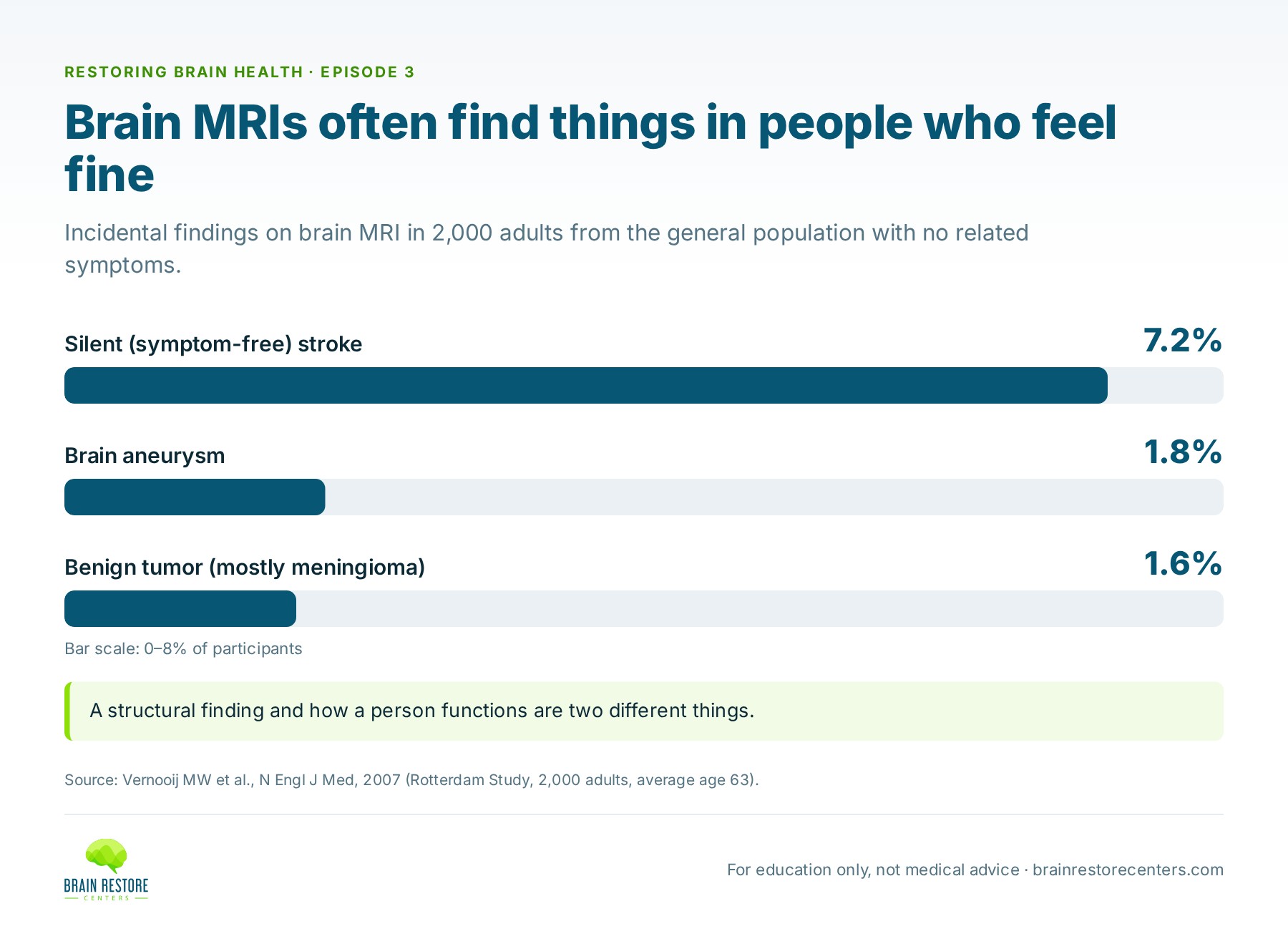
Research backs this up on a large scale. When researchers in the Rotterdam Study ran brain MRIs on 2,000 adults from the general population (average age 63), they found silent brain findings in a surprising number of people: 7.2% had a small stroke that had never caused symptoms, 1.8% had a brain aneurysm, and 1.6% had a benign tumor, mostly meningiomas. None of these people had come in because of symptoms. A structural finding and how a person functions are two different things.
Can your brain look normal on imaging but still not work normally?
This is the key point for anyone with a clear brain MRI or CT and ongoing symptoms. “Just because they’re structurally okay also doesn’t mean they’re functionally okay,” Dr. Webb says. Even someone with a very small bleed who seems completely normal can go on to develop concussion symptoms, or symptoms so mild they don’t notice them at first.
The largest U.S. brain injury study, called TRACK-TBI, has followed thousands of people seen in trauma centers. One analysis looked at 991 adults who had a mild head injury, were fully alert in the emergency room, and had a CT that showed no injury. At six months, 56% of those followed up had not fully recovered, and most of them said they hadn’t gotten back to their pre-injury life. The authors recommended a follow-up visit about two weeks after injury so these people don’t fall through the cracks.
We go deeper on this, including what MRI can find that CT misses and what it means for kids, in can a normal brain MRI or CT miss a brain injury? If you’re new to the idea, Episode 1 covers why your tests can be normal when you don’t feel normal.
Don’t functional MRI and DTI measure how the brain works?
People ask this a lot, and the names don’t help. A functional MRI tracks changes in blood oxygen while you do a task, like reading, to show which areas are working harder during that task. Its main clinical use is surgical planning: finding exactly where a person’s language or movement areas sit before removing a nearby tumor. Diffusion tensor imaging (DTI) follows the movement of water to map the brain’s white-matter wiring. It also mainly supports surgical planning, and it can show more serious axonal injury.
“These aren’t functional tests. They’re not looking at the function of the brain.”
— Dr. Sharon Webb, on Restoring Brain Health, Episode 3
Both are found mostly at academic medical centers. For how each one works and what the research shows, read does a functional MRI measure how well your brain works?
Brain imaging vs. functional assessment: side by side
| Standard CT / MRI | Functional MRI (fMRI) | Diffusion tensor imaging (DTI) | Functional assessment battery | |
|---|---|---|---|---|
| The question it answers | Is there a structural problem, like a bleed, fracture, stroke or mass? | Which areas of the brain get more blood flow during a specific task? | Where do the brain’s white-matter pathways run, and is there visible injury to them? | How well are the brain’s networks working right now? |
| What it actually measures | Anatomy: bone, tissue, blood vessels, swelling | Changes in blood flow while you do a task, such as reading or moving a hand | The direction and integrity of white-matter tracts | Brainwave patterns (qEEG brain map), balance, eye movements, memory and thinking, a hands-on neurological exam, and your history |
| Main use | Ruling out danger after an injury or new symptoms | Mostly surgical planning: mapping language and motor areas near a tumor | Surgical planning, and sometimes showing more serious axonal injury | Getting a functional baseline when symptoms continue, and measuring again later |
| Does it measure how well the brain is working? | No | Not in the everyday sense. It shows where activity happens during a task | No | Yes. That’s its purpose |
| Where you can get it | Hospitals and imaging centers | Mostly academic medical centers | Mostly academic medical centers | A trained clinician’s office |
| The analogy | A photo | A highlighted map of one task | A wiring diagram | A video |
Each tool answers a different question. Imaging is essential when your doctor suspects a structural problem; a functional assessment adds a different kind of information alongside it.
How is brain function actually measured?
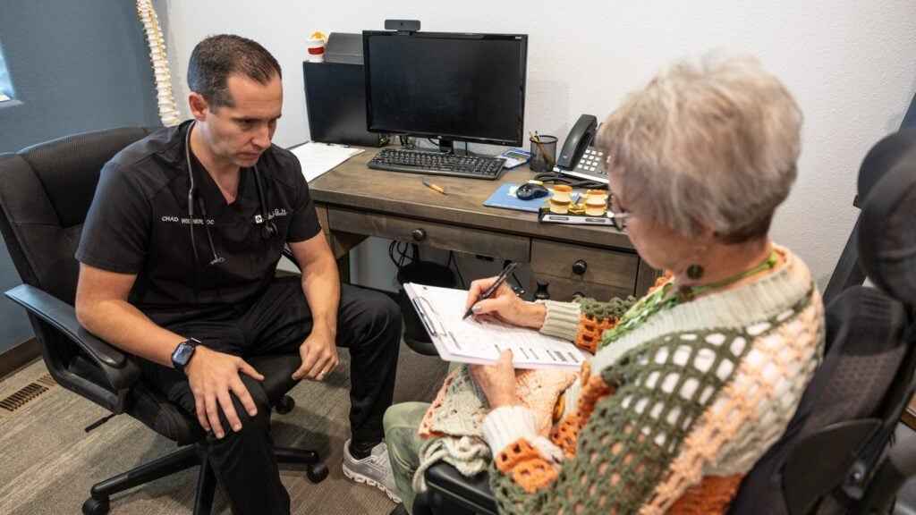
Dr. Trevor Berry, whom Dr. Woolner describes as an integrative functional neurologist, starts where every good clinician starts: a careful history and a bedside neurological exam. A good history already points somewhere. Dizziness and balance trouble may suggest the brain stem; other patterns point toward the frontal or parietal lobes. From there, objective tests confirm or challenge that first impression:
- A qEEG brain map. Sensors on the scalp listen to brainwave activity, and the results are compared with normative data from large groups of people to show areas that may be overactive or underactive.
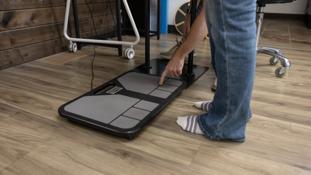
- Computerized balance testing. A force plate measures exactly how a person sways, forward, back or to one side. Dr. Berry calls it one of the most sensitive tests you can do on the nervous system.
- Eye movement, coordination and strength checks, such as finger-to-nose testing, fast eye movements and grip strength.
- A memory and thinking screen and a detailed symptom history.
No single result is the answer. When findings line up across several tests, the picture becomes much clearer. At Brain Restore Centers, when that picture shows areas of the brain not working together as well as they should, we call it network interference. Network interference is what we call it when testing shows that different areas and networks of the brain aren’t functioning and working together as well as they should. It’s something we measure, not a diagnosis.
“We are not putting all of our eggs in one singular basket.”
— Dr. Chad Woolner, on Restoring Brain Health, Episode 3
Why several tests beat one, with the research on sensitivity and test batteries, is covered in why one brain test isn’t enough.
Why does access to functional testing matter?
Dr. Berry loves advanced tests like SPECT and DTI, but as he points out, many people only get them after being admitted to a hospital for something serious. And when Dr. Woolner asked Dr. Webb how long a new patient waits to see her, the answer was “multiple months.” That isn’t because anyone is uncaring. The system is stretched.
Functional testing doesn’t require a multimillion-dollar machine. In the hands of a trained clinician, these relatively low-tech tests can show clear functional patterns now, and give you a baseline you can measure again later. That’s why Brain Restore Centers is building a network of centers across the country, so this kind of look at the brain is available close to home instead of only at a few academic hospitals.
Who is this for?
- Adults noticing memory or thinking changes who were told their imaging looks fine for their age and to come back in a year.
- People recovering from a concussion or brain injury whose CT or MRI was clear but who still have headaches, fog, dizziness or mood changes.
- Parents of kids recovering from a concussion who want to understand how their child’s brain is working, not just whether something is visible on a picture.
- Anyone who wants a baseline of their brain health to track over time.
Frequently asked questions
Is a brain MRI the same as a functional brain assessment?
No. A brain MRI shows structure. A functional assessment looks at how the brain is performing, using tools like a qEEG brain map, balance testing and a neurological exam. They answer different questions and work well together.
Is a CT or an MRI better after a head injury?
They do different jobs. CT is fast and excellent for finding bleeding or fractures in an emergency. MRI shows more detail and can find smaller injuries. Your doctor decides which you need based on your injury and symptoms.
If my imaging was clear, do I still need to follow up with my doctor?
Yes. Keep your follow-up appointments, and tell your doctor if symptoms change. Functional testing adds information alongside your doctor’s care; it doesn’t replace it.
Can one test tell me what’s going on?
Rarely. Even very sensitive tests have limits. That’s why a battery of tests, read together with your history and exam, gives a more reliable picture than any single result.
Studies referenced
- Vernooij MW, Ikram MA, Tanghe HL, et al. Incidental findings on brain MRI in the general population. N Engl J Med. 2007;357(18):1821-1828. https://pubmed.ncbi.nlm.nih.gov/17978290/
- Madhok DY, Rodriguez RM, Barber J, et al. Outcomes in patients with mild traumatic brain injury without acute intracranial traumatic injury. JAMA Netw Open. 2022;5(8):e2223245. https://pubmed.ncbi.nlm.nih.gov/35976650/
Find out how your brain is working right now
The first step is a Complimentary Brain Scan at a Brain Restore Center near you: a functional look at how your brain is working today. No pressure, no assumptions. If it looks like we may be able to help, we’ll talk about next best steps.
This article is for education and is not medical advice. It does not replace care from your own doctor. If you have sudden or severe symptoms, such as a sudden severe headache, new weakness or numbness, trouble speaking, or confusion, call 911 or go to the nearest emergency room.