The short answer
Because every test has limits. Some tests are very good at catching a problem but can’t say what it is; others are precise but miss a lot. In one study of concussed athletes, single tests caught between 44% and 79% of injuries, while the full battery caught more than 90%. When a history, a neurological exam and several objective tests point the same way, you can be far more confident about what’s going on.
It’s tempting to want one device that gives the whole answer. As Dr. Chad Woolner joked on Episode 3 of Restoring Brain Health, that would take a “Star Trek” wand you wave over someone. In real life, good clinicians build the picture in layers. Here’s the science behind why.
What do sensitivity and specificity mean?
These are the two words Dr. Woolner says he brings up with patients “time and time again.” They’re also the two basic measures of any medical test, as a classic BMJ series on diagnostic tests explains:
- Sensitivity is how good a test is at catching a problem when it’s really there. A highly sensitive test rarely misses.
- Specificity is how good a test is at saying “no problem” when there really isn’t one. A highly specific test rarely raises a false alarm.
Think of a smoke alarm. A very sensitive alarm goes off for every real fire, but also for burnt toast. A very specific alarm never goes off for toast, but might be slow to catch a small fire. Most tests sit somewhere in between, and making one better often makes the other worse.
“In an ideal world, we would have a belt of tests, so to speak, that are each 100% sensitive and 100% specific,” Dr. Woolner says. “That’s not reality, though.” There will always be some uncertainty. Combining tests is how clinicians make up for it.
What does research show about using a battery of tests?
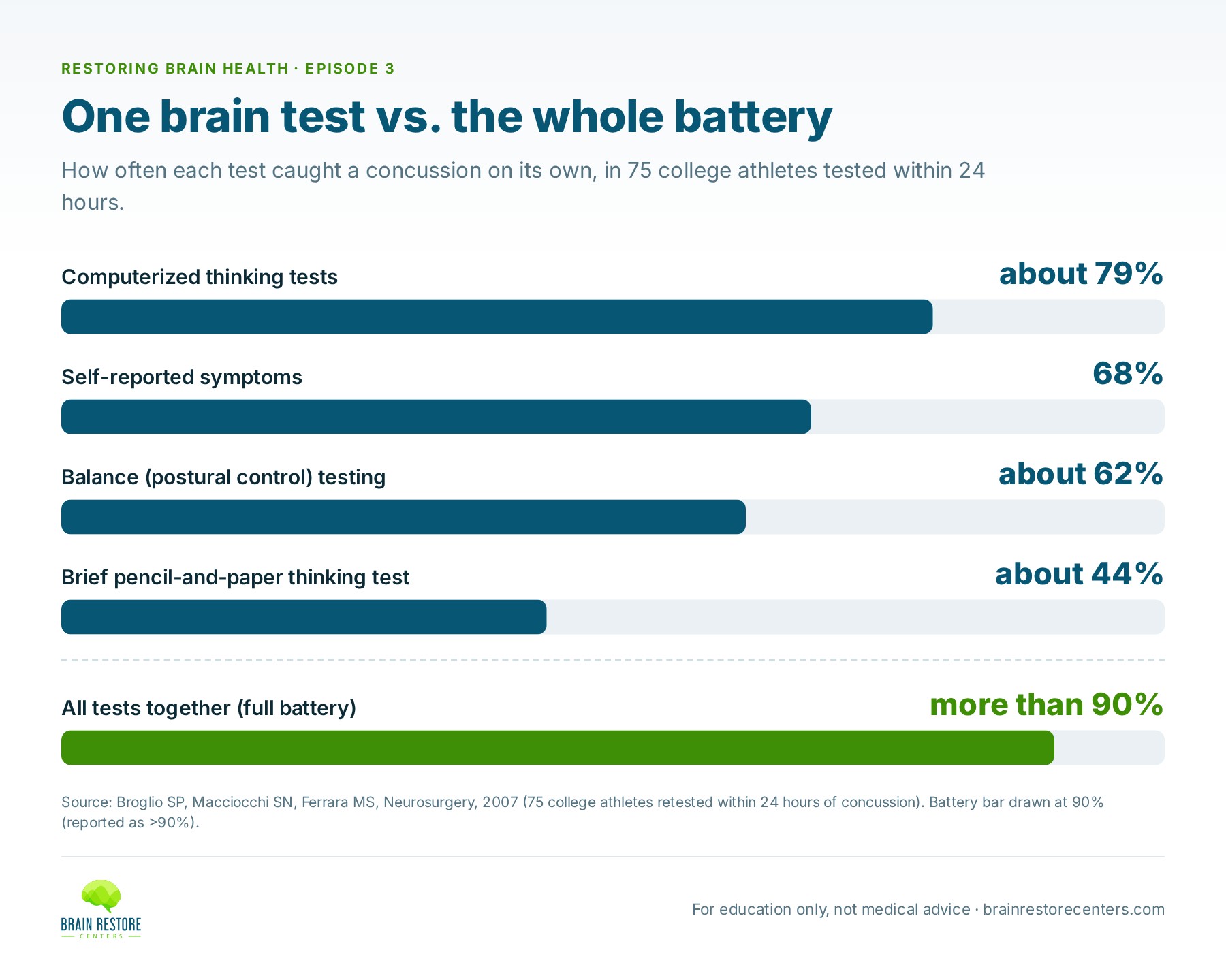
Concussion research gives a clear example. Researchers at the University of Illinois tested college athletes before their seasons, then retested 75 athletes within 24 hours of a diagnosed concussion. They compared how often each test caught the injury on its own:
- Computerized thinking tests: about 79%
- Self-reported symptoms: 68%
- Balance (postural control) testing: about 62%
- A brief pencil-and-paper thinking test: about 44%
- The full battery together: more than 90%
Each test looked at a different part of brain function, and each missed some injured athletes. Together, they caught almost all of them. The authors concluded that concussion should be approached through a multifaceted assessment that looks at different aspects of function. That’s the same logic behind Brain Restore Centers’ approach.
“We are not putting all of our eggs in one singular basket.”
— Dr. Chad Woolner, on Restoring Brain Health, Episode 3
Why can’t even a very sensitive test diagnose on its own?
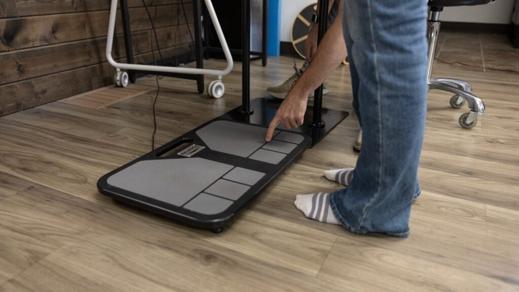
Dr. Trevor Berry, whom Dr. Woolner describes as an integrative functional neurologist, points to computerized balance testing. In computerized dynamic posturography, you stand on a force plate that measures tiny shifts in pressure while the platform or your surroundings move. It maps exactly how you sway, forward and back or side to side, and how well you use your eyes, your inner ear and the feeling in your feet to stay upright.
It’s a sensitive window into the nervous system. In a study of 36 college athletes with concussion, balance testing on this kind of system picked up clear deficits the day after injury, which the researchers linked to the brain not using information from the visual and vestibular (inner ear) systems effectively.
Yet Dr. Berry is clear about its limits. Balance patterns may look more typical of a brain injury or of a condition like Parkinson’s, “but you’re not going to diagnose from that test.” A sensitive test tells you something is off and where to look next. It doesn’t tell you the whole story.
Why does a finding only matter if it fits the person?
Dr. Sharon Webb, a neurosurgeon, gave an example from spine surgery. Someone comes in with back pain running down the leg. The MRI shows a disc bulge near the nerve, but it’s “not very convincing.” The symptoms come and go, and on the day of the exam there’s no weakness. So she looks for more confirmation, such as how the person responds to a targeted injection at that level, before deciding the bulge is really the problem.
There’s a good reason for that caution. A Mayo Clinic review of 33 studies covering 3,110 people with no back pain found disc bulges on imaging in 30% of 20-year-olds and 84% of 80-year-olds. The authors wrote that these findings must be interpreted in the context of the patient’s clinical condition.
“You can have a finding, but the finding’s only as good as it correlates with all the other data you have and the history and the patient’s exam itself.”
— Dr. Sharon Webb, on Restoring Brain Health, Episode 3
That’s true, she adds, “whether it’s conventional medicine or functional medicine.”
How do layers of testing point to the right area of the brain?
Dr. Berry walks through it step by step. If someone describes dizziness and balance trouble, his first thought may be the brain stem. Then he looks for confirmation:
- History. What are the symptoms, when did they start, what makes them better or worse?
- Bedside exam. A finger-to-nose test may show dysmetria, a slight overshoot that can point toward the cerebellum. He doesn’t stop at one sign; he runs more cerebellar tests and checks the rest of the nervous system.
- Computerized balance testing to measure sway patterns precisely.
- A qEEG brain map to see how brainwave activity compares with normative data.
- Eye movements and grip strength as further checks.
“The more layers of confirmation, the more I can be confident,” he says. At Brain Restore Centers, when the layers show networks not working together as they should, we call it network interference. Network interference is what we call it when testing shows that different areas and networks of the brain aren’t functioning and working together as well as they should. It’s something we measure, not a diagnosis.
Where does a qEEG brain map fit in?
Dr. Berry calls qEEG “one of our best windows” into brain function. Sensors on the scalp listen to brainwave activity across different frequencies. The “quantitative” part means a computer compares the recording with normative data collected from large numbers of people.
The idea goes back decades. In 1977, a team led by E. Roy John published “Neurometrics” in Science, laying out the approach of measuring brain electrical activity and comparing it statistically with norms. Today, as Dr. Berry explains, comparisons drawn from large databases show recognizable patterns. Read on its own, a qEEG is one layer. Read alongside the exam, balance testing and history, it helps point to where the brain’s communication is off.
Why measure again?
Objective tests give you a baseline. Dr. Berry and Dr. Webb both stress reassessment: measure, then measure again later to see what has actually changed, instead of relying on guesswork or memory.
How to use this as a patient
- Ask what question each test you’re offered is meant to answer.
- Be cautious about any single gadget presented as the whole answer. As Dr. Woolner notes, there’s a new brain gadget ad on social media almost every day.
- Bring your full history; it’s part of the data.
- Ask whether results will be measured again over time.
For how imaging fits into all of this, read the photo and the video: what brain imaging shows, and what it can’t, or see what functional MRI and DTI really show.
Studies referenced
- Altman DG, Bland JM. Diagnostic tests 1: sensitivity and specificity. BMJ. 1994;308(6943):1552. https://pubmed.ncbi.nlm.nih.gov/8019315/
- Broglio SP, Macciocchi SN, Ferrara MS. Sensitivity of the concussion assessment battery. Neurosurgery. 2007;60(6):1050-1057. https://pubmed.ncbi.nlm.nih.gov/17538379/
- Guskiewicz KM, Ross SE, Marshall SW. Postural stability and neuropsychological deficits after concussion in collegiate athletes. J Athl Train. 2001;36(3):263-273. https://pubmed.ncbi.nlm.nih.gov/12937495/
- Brinjikji W, Luetmer PH, Comstock B, et al. Systematic literature review of imaging features of spinal degeneration in asymptomatic populations. AJNR Am J Neuroradiol. 2015;36(4):811-816. https://pubmed.ncbi.nlm.nih.gov/25430861/
- John ER, Karmel BZ, Corning WC, et al. Neurometrics. Science. 1977;196(4297):1393-1410. https://pubmed.ncbi.nlm.nih.gov/867036/
Watch the full conversation: Restoring Brain Health, Episode 3.
Find out how your brain is working right now
The first step is a Complimentary Brain Scan at a Brain Restore Center near you: a functional look at how your brain is working today. No pressure, no assumptions. If it looks like we may be able to help, we’ll talk about next best steps.
This article is for education and is not medical advice. It does not replace care from your own doctor. If you have sudden or severe symptoms, such as a sudden severe headache, new weakness or numbness, trouble speaking, or confusion, call 911 or go to the nearest emergency room.