The short answer
A normal CT or MRI means no dangerous structural problem was found, such as bleeding, swelling, a fracture or a mass. That’s real good news. But those tests take a picture of the brain’s structure. They were not built to measure how well the brain is working. You can have a clear brain MRI or CT and still have a real problem with function. Both can be true at the same time.
You know something is off. Maybe it’s brain fog, words that won’t come, or reading the same paragraph four times. Maybe it’s a parent who keeps repeating the same stories, or a teenager who hasn’t been the same since a hit on the field. So you see a doctor. The imaging comes back clear. The labs are fine. And you leave wondering: is this all in my head?
It isn’t. This guide explains why normal brain imaging and real symptoms can exist side by side, what large research studies have found about people in exactly this spot, and what it looks like to measure how the brain is working, not just how it looks.
This article is based on Episode 1 of Restoring Brain Health, the Brain Restore Centers podcast with Dr. Chad Woolner, Dr. Sharon Webb and Dr. Trevor Berry.
Prefer to listen? Restoring Brain Health is on every major podcast app:
Listen on SpotifyListen on Apple PodcastsSubscribe on YouTube
Key takeaways
- A normal CT or MRI rules out structural problems large enough to see. That matters, and it’s good news.
- Imaging was never designed to measure function: how well brain regions communicate and work together.
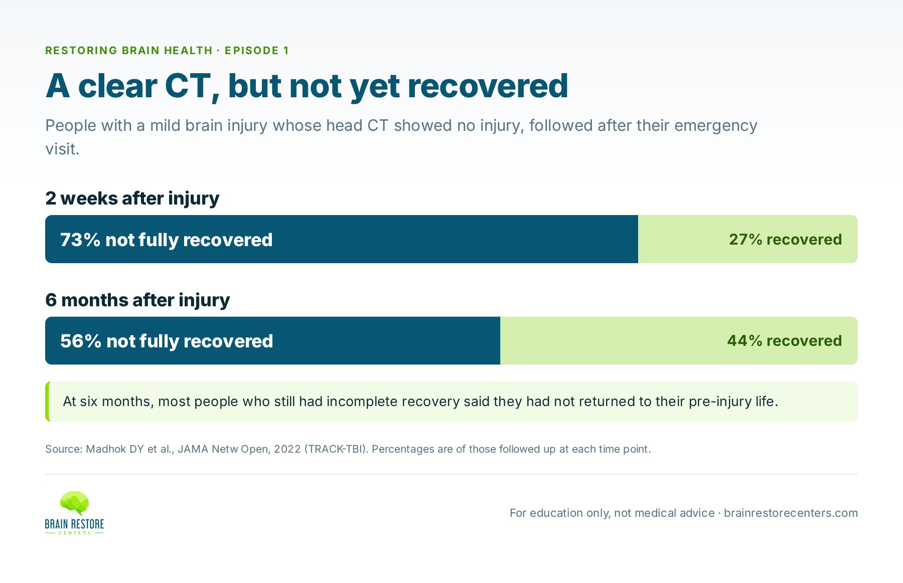
- In a large U.S. study of people with a mild brain injury and a clear CT, 56% had not fully recovered six months later.
- Research tools show that the brain’s networks can change how they work even when people still perform tasks correctly.
- Function can be measured with tools like qEEG brain mapping, balance testing, memory and thinking screens and a neurological exam.
- Your history matters. In classic studies, the patient’s own story led to the right diagnosis most of the time.
What is a brain MRI or CT actually looking for?
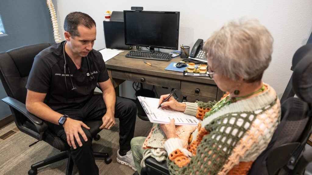
When someone has new neurological symptoms, especially after a fall or an injury, the first job is to rule out anything dangerous or fixable. On Episode 1, Dr. Sharon Webb, a dual-trained neurosurgeon, describes what that imaging is searching for: a skull fracture, bleeding inside the skull, swelling of the brain, or “something structural that’s going to explain why you’re having these issues.”
Labs play a similar role. As she points out, a fall might trace back to something like a urinary tract infection that caused confusion. The goal is to find “the severe or structural issues that can be fixed or even need to be followed, but certainly need to be known about.”
That job is essential, and imaging does it well. When the imaging is clear, it is real relief. As Dr. Chad Woolner puts it on the show, the good news is “you’re not dealing with a tumor, you’re not dealing with a brain bleed.” But he also names the catch: a clear brain image “doesn’t necessarily tell us what is going on.”
The photo and the video: why structure isn’t the same as function
Dr. Trevor Berry offers the analogy at the heart of this episode.
“Those tests do a good job of taking a picture, but your brain is creating a video.”
— Dr. Trevor Berry, on Restoring Brain Health, Episode 1
He gives a vivid example he uses with patients. If he put both arms into an MRI tube, the report might say the muscles look balanced and equal. “Well, how do you know one’s not paralyzed?” The structure can look perfect while the function is not.
The same is true for the brain. Your brain is a communication network. Billions of nerve cells send signals across regions that have to talk to each other in the right order, at the right speed, and at the right strength. A photo can show that all the parts are present. It can’t show whether the conversation between them is running smoothly.
Structural imaging vs. functional assessment, side by side
| Structural imaging (CT, MRI) | Functional assessment | |
|---|---|---|
| The question it answers | Is something structurally wrong or dangerous? | How well is the brain working right now? |
| What it looks at | Bone, tissue, bleeding, swelling, masses | Brainwave activity, balance, memory and thinking, reflexes and eye movements |
| Analogy | A photo | A video |
| What “normal” means | No dangerous structural problem was found. That’s good news. | Results compared with people of the same age, so you can see where function sits |
| What it can’t tell you | How well different areas are communicating and working together | It doesn’t replace imaging when your doctor suspects a structural problem |
| When it’s used | After an injury, or for sudden, severe or worsening symptoms. Your doctor decides. | When symptoms continue and structural tests are clear, or to set a baseline |
The two answer different questions. Most people who are struggling are best served by both.
What happens to people whose brain CT is normal after an injury?
This is one of the clearest windows into the gap between structure and function. TRACK-TBI is a large U.S. research program that follows people seen in emergency departments for brain injury at 18 major trauma centers. In 2022, researchers looked at 991 adults who had a mild brain injury, were fully alert in the ER, and had a head CT showing no injury.1
These were, by the usual standard, the “all clear” patients. Yet at two weeks, 73% of those followed up had not fully recovered. At six months, 56% still had incomplete recovery, and most of them said they had not returned to their pre-injury life.
That doesn’t mean the CT was wrong. It did its job: no bleeding, no fracture, nothing that needed surgery. It means a clear CT and a full recovery are not the same thing. For a lot of people, the real question starts after the imaging comes back normal.
Can the brain have a real problem that doesn’t show up on imaging?
Yes. Dr. Webb explains it plainly on the episode. Standard imaging picks up “large enough structural damage,” while “small microscopic neurological damage” can still affect how the brain works. “It’s at the cellular level,” she says. “So you’re going to need different tools that we just don’t really use in conventional medicine.”
Research backs this up from two directions. First, even within imaging, the tool matters. In an earlier TRACK-TBI study, 27% of people with mild brain injury whose admission CT was normal showed findings on a brain MRI done about two weeks later.2 Finer tools see finer things.
Second, scientists can study brain networks directly with advanced research methods. In a study published in the journal Brain, researchers at Imperial College London used functional MRI to compare people living with the long-term effects of brain injury against healthy people of the same age.3 The patients answered a simple task correctly, but more slowly and less consistently. Their brain activity showed changes in a key network, in a pattern the authors described as consistent with an increased mental workload. In other words, their brains were getting the job done, but working harder to do it.
That is exactly what so many people describe: “I can still do it, but it’s exhausting.” Advanced research scanners like that aren’t part of a routine clinic visit. But they show that changes in how the brain’s networks work are real and measurable.
What is network interference?
On the podcast, the hosts use one term for this idea. Network interference is what we call it when testing shows that different areas and networks of the brain aren’t functioning and working together as well as they should. It’s something we measure, not a diagnosis.
Dr. Berry describes what testing can pick up: whether “your brain is working too hard or not working hard enough, it maybe doesn’t have enough energy, it’s inflamed or not,” and whether “those signals and those communication networks are being compromised in some way, shape, or form.”
How do you measure how the brain is working?
No single test tells the whole story, so a functional assessment looks from several angles:
- A qEEG brain map. Sensors on the scalp record the brain’s electrical activity, and software compares the patterns with a database of people the same age.
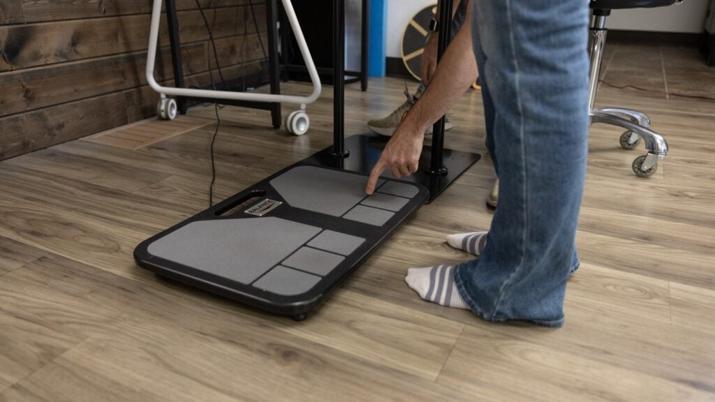
- Computerized balance testing. Standing still is a team effort between your eyes, inner ear and body sense. A force plate measures how well the brain is coordinating them.
- A standardized memory and thinking screen that measures attention, recall and processing speed.
- A hands-on neurological exam: reflexes, strength, eye movements, coordination and sensation.
- Your history and symptom picture, read carefully, because your story is data too.
Together they show patterns no single test could. And because they can be repeated, they give you a baseline to compare against later. Our spokes go deeper on each angle: why you can feel off when tests are normal, exactly what a normal MRI or CT rules out, and what to do next.
Why listening to the patient still comes first
Dr. Webb is emphatic on this point. A history and physical exam are what tell a doctor an image is needed in the first place.
“We don’t treat an image, we treat a patient.”
— Dr. Sharon Webb, on Restoring Brain Health, Episode 1
The research agrees. In a classic study in the British Medical Journal, doctors reached the diagnosis that turned out to be correct after reading the referral letter and taking the history in 66 of 80 new patients.4 The exam and lab tests each added the answer in only seven more. Lab work and imaging are vital for ruling things out. But your description of what has changed is often the most important information in the room.
Who this is for
- Adults worried about memory, or families watching a loved one change, who were told to come back in six months.
- People after a concussion or brain injury whose brain MRI or CT was clear but who still aren’t themselves.
- Parents of kids and teens still struggling weeks after a concussion.
- Anyone whose brain just isn’t working the way it used to and who wants information instead of waiting.
As Dr. Woolner says on the show, the first message is simple: you’re not crazy. “Watch and wait” leaves people with nothing to measure and nothing to do. A functional baseline gives you something concrete to work from.
Frequently asked questions
My brain MRI was normal. Do I need another one?
That’s a question for your doctor, and it depends on your symptoms and history. A repeat MRI answers the same structural question again. If you want to know how well your brain is working, that’s a different kind of test.
Does a normal brain MRI mean it’s all in my head?
No. A normal brain MRI or CT means no dangerous structural problem was found. It doesn’t measure function, so it can’t rule out that your brain is working harder than it should.
Is functional testing a replacement for my doctor’s tests?
No. It answers a different question. Keep your follow-up appointments. Functional testing adds information alongside them.
Is a Complimentary Brain Scan the same as an MRI?
No. It’s a functional screening that looks at how your brain is working today. It doesn’t diagnose, and it doesn’t replace imaging your doctor orders.
Studies referenced
- Madhok DY, Rodriguez RM, Barber J, et al.; TRACK-TBI Investigators. Outcomes in patients with mild traumatic brain injury without acute intracranial traumatic injury. JAMA Netw Open. 2022;5(8):e2223245. PubMed
- Yuh EL, Mukherjee P, Lingsma HF, et al.; TRACK-TBI Investigators. Magnetic resonance imaging improves 3-month outcome prediction in mild traumatic brain injury. Ann Neurol. 2013;73(2):224–235. PubMed
- Sharp DJ, Beckmann CF, Greenwood R, et al. Default mode network functional and structural connectivity after traumatic brain injury. Brain. 2011;134(Pt 8):2233–2247. PubMed
- Hampton JR, Harrison MJ, Mitchell JR, Prichard JS, Seymour C. Relative contributions of history-taking, physical examination, and laboratory investigation to diagnosis and management of medical outpatients. Br Med J. 1975;2(5969):486–489. PubMed
Find out how your brain is working right now
The first step is a Complimentary Brain Scan at a Brain Restore Center near you: a functional look at how your brain is working today. No pressure, no assumptions. If it looks like we may be able to help, we’ll talk about next best steps.
This article is for education and is not medical advice. It does not replace care from your own doctor. If you have sudden or severe symptoms, such as a sudden severe headache, new weakness or numbness, trouble speaking, or confusion, call 911 or go to the nearest emergency room.