The short answer
A normal CT or brain MRI rules out structural problems large enough to see, such as a skull fracture, bleeding, swelling, a stroke or a mass. That’s important news. What it doesn’t measure is function: how well different areas of your brain are working and communicating. That takes different tools.
“Your MRI is normal” can feel like both relief and a dead end. This guide explains how brain imaging works, what a clear result really tells you, and why, as Dr. Chad Woolner puts it on Episode 1 of Restoring Brain Health, a clear brain image “doesn’t necessarily tell us what is going on.”
How do brain CT and MRI work?
Both create detailed pictures of the inside of your head, but in different ways.
A CT (computed tomography) uses X-rays taken from many angles. A computer combines them into slices. Dense things like bone look bright, and fresh blood stands out clearly. CT is fast, which is why it is usually the first test after a head injury in the emergency room.
A brain MRI (magnetic resonance imaging) uses a powerful magnet and radio waves instead of X-rays. It picks up signals from water in your tissues, which lets it show soft brain tissue in much finer detail than CT.
| Brain CT | Brain MRI | |
|---|---|---|
| How it works | X-rays taken from many angles, combined by a computer into cross-section images | A strong magnet and radio waves detect signals from water in tissue |
| Best at | Fast checks for fresh bleeding, skull fractures and swelling. Often the first test in the ER. | Fine detail of brain tissue, small areas of injury, strokes, masses and white matter |
| Typical time | Minutes | Often 30 to 60 minutes |
| What neither shows | How well brain regions are communicating | How well brain regions are communicating |
Both are structural tests. Your doctor chooses which one fits your situation.
What a normal brain MRI or CT rules out
According to neurosurgeon Dr. Sharon Webb, imaging after symptoms or an injury is looking for anything dangerous or fixable, including:
- A skull fracture
- Bleeding inside the skull (intracranial hemorrhage)
- Swelling of the brain (cerebral edema)
- A mass or tumor
- Other structural changes that need surgery or close follow-up
If those come back clear, that is genuine relief. “You’re not dealing with a tumor, you’re not dealing with a brain bleed,” as Dr. Woolner says. Take that good news seriously.
Does a normal CT mean the brain wasn’t injured?
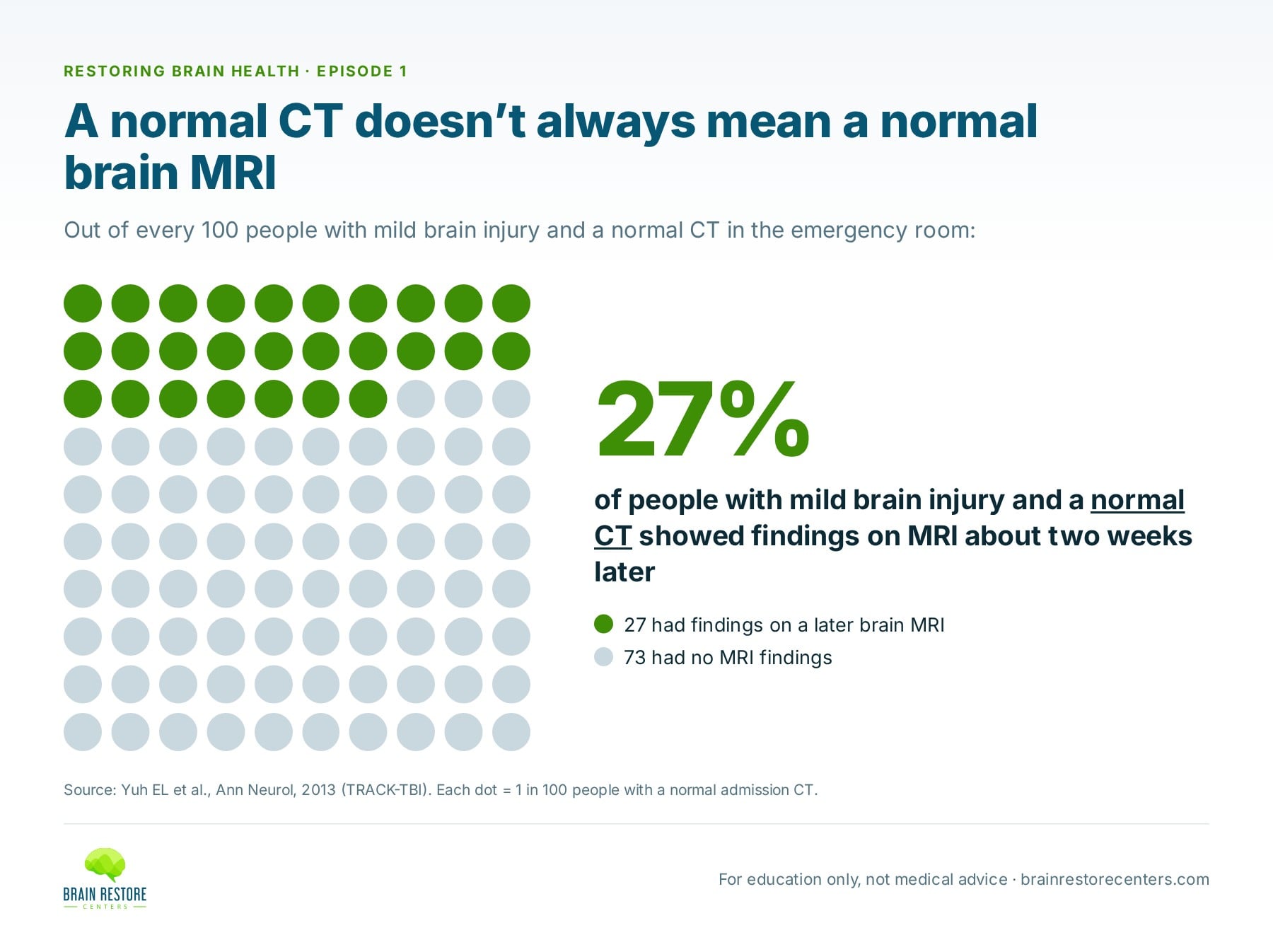
Not necessarily. Different tools see different levels of detail. In a study of people seen in emergency departments for mild brain injury, researchers repeated imaging with an MRI about 12 days after the injury. 27% of people whose CT had been normal showed findings on MRI.1 Some of those findings were linked to a slower recovery at three months.
That isn’t a knock on CT. It’s the right first test for finding emergencies fast. It simply shows a principle: a normal result means “nothing found with this tool,” not “nothing there.”
What can’t a normal brain MRI or CT tell you?
- How well different regions of the brain are communicating
- Whether some areas are working too hard or not hard enough
- How your balance, memory and processing speed are performing
- Changes at the cellular level that are too small to see
Dr. Trevor Berry’s analogy captures it: imaging takes a picture, but “your brain is creating a video.” A picture can show every part in place without showing whether the parts are working together.
What tests look at brain function instead?
Some functional tools are mainly used in research, like functional MRI, which tracks blood flow linked to brain activity. Others are practical in a clinic. One of the most established is EEG.
qEEG: measuring the brain’s electrical activity
Your brain cells communicate with tiny electrical signals. An EEG records that activity through sensors on the scalp. A quantitative EEG (qEEG) goes a step further: software breaks the signal into its frequencies, measures how regions are timed with each other, and compares the results with a database of people the same age. The result is often called a brain map.
This approach has decades of research behind it. In a 1989 study, Robert Thatcher and colleagues recorded EEGs from 608 people with mild head injury and 108 healthy people.2 An analysis of the EEG patterns separated the two groups with high accuracy, above 90% in several independent test groups. The differences they found were exactly the kind of thing a structural image isn’t built to see: changes in how regions were timed and connected with each other, and reduced alpha activity at the back of the brain.
That is why qEEG is often described as showing what MRIs miss.
Balance, memory and the neurological exam
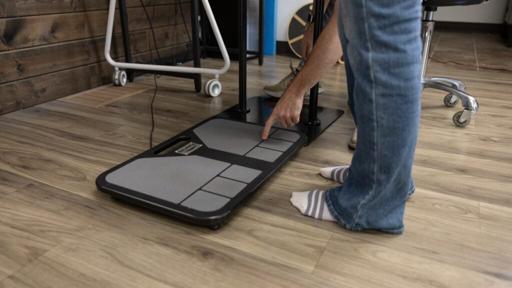
Balance testing, memory and thinking screens, and a hands-on neurological exam each add a different view. We cover them in what to do after you’re told everything looks normal.
At Brain Restore Centers, when testing shows that areas of the brain aren’t working together as well as they should, we call it network interference. It’s something we measure, not a diagnosis.
Questions to ask your doctor about your imaging
- What was this imaging looking for?
- Was it a CT or an MRI, and why that one?
- Did anything need follow-up, even if it’s not related to my symptoms?
- What changes in my symptoms would mean I should come back sooner?
- Is it reasonable for me to look at how my brain is functioning in the meantime?
“We don’t treat an image, we treat a patient.”
— Dr. Sharon Webb, on Restoring Brain Health, Episode 1
Want the full picture? See structural imaging vs. functional assessment, side by side, or read five reasons you can feel off when your tests are normal.
Studies referenced
- Yuh EL, Mukherjee P, Lingsma HF, et al.; TRACK-TBI Investigators. Magnetic resonance imaging improves 3-month outcome prediction in mild traumatic brain injury. Ann Neurol. 2013;73(2):224–235. PubMed
- Thatcher RW, Walker RA, Gerson I, Geisler FH. EEG discriminant analyses of mild head trauma. Electroencephalogr Clin Neurophysiol. 1989;73(2):94–106. PubMed
Watch the full conversation: Restoring Brain Health, Episode 1.
Find out how your brain is working right now
The first step is a Complimentary Brain Scan at a Brain Restore Center near you: a functional look at how your brain is working today. No pressure, no assumptions. If it looks like we may be able to help, we’ll talk about next best steps.
This article is for education and is not medical advice. It does not replace care from your own doctor. If you have sudden or severe symptoms, such as a sudden severe headache, new weakness or numbness, trouble speaking, or confusion, call 911 or go to the nearest emergency room.